Showing posts with label chart. Show all posts

Monday, November 29, 2021

Petri Dish Bacteria Identification Chart

For example the Microbiologist might. Beta-hemolysis on media with blood sheep horse Yellow-pigmented colonies.

Mold Petri Dish Identification Chart Page 1 Line 17qq Com

L Lunger Nature Inspiration.

Petri dish bacteria identification chart. I have developed software applications that will enable users to identify the organisms based on the results of their tests. Be sure to label the bottom of the petri dish rather than the lid as the lid will turn causing your label to be over the wrong part of your dish. Production of coagulase free and cell-bound coagulase Lithium chloride and potassium tellurite tolerant.

It can be used to help to identify them. Grains with and without surface sterilization were transferred separately to pre-sterilized petri dish humid chambers under aseptic conditions. Touch plates showing a variety of bacteria found on the fingertips.

Lecithinase production and lipase activity. Place a microscope slide inside a petri dish. The phenazine null strain Δphz started to wrinkle on day 2 the wild type wt wrinkled on day 3 and the soxR and mexGHI-opmD deletion strains wrinkled on day 5 whereas a pyocyanin overproducer DKN370 remained smooth and white after 6 days.

A colony is defined as a visible mass of microorganisms all originating from a single mother cell therefore a colony constitutes a clone of bacteria all genetically alike. Different agar plates are available from EMSL Analytical Inc depending on the types of fungi or bacteria to be identified. Colony morphology is a method that scientists use to describe the characteristics of an individual colony of bacteria growing on agar in a Petri dish.

The fungi or bacteria are counted and identified. The petri dishes were incubated for 5 days at 281 C in an incubator with a 12-h light cycle. Keep the petri dish cover available.

Be careful not to. Microbiologists use a number of terms to describe the different appearances of bacteria. Being able to visibly differentiate bacteria based on the appearance of their colonies is a crude but essential first step in isolating the different types of bacteria in the sample.

It is useful for bacteria identification. First and foremost it can be done visually by looking at the colonies ie the cultural characteristics and noting the following. We should examine the bacteria colonies in the closed petri dish for safety.

Bacteria grow on solid media as colonies. Coccobacilli elongated coccal form. Measuring the diameter of the colony.

EMSL VP-400 Microbial Sampler Product ID 8709001. Bacilli rod-shaped coccus pl. Each grain sample 200 grains treatment 1 was examined to identify fungi up to the species level.

Colony morphology is a method that scientists use to describe the characteristics of an individual colony of bacteria growing on agar in a Petri dish. This method commonly uses an Andersen N-6 type impactor eg. NaCl tolerant 75 mannitol fermentation.

Gram-positive or Gram-negative coccus or bacillus etc. Once you have introduced the bacteria you should replace the lid on the Petri dish and seal it with some parafilm or Saran wrap. The Petri dishes are incubated in the laboratory so the organisms impacted on the plate can grow.

The software uses probability matrix for identification and the results are expressed in percentage probabilities. Armed with cotton swabs and Petri dishes full of nutient agar students head out of the lab to see what lives on surfaces they encounter everyday. Make sure to label each Petri dish with the source of the bacteria.

For example circular filamentous etc. Cocci sounds like cox-eye spherical spirillum pl. The matrix table can also be used to view the properties of these organisms and to compare their properties.

Elevation - What is the cross sectional shape of the colony. Using a sterile inoculating loop or wooden applicator stick collect a small amount of organism from a well-isolated 18- to 24-hour colony and place it onto the microscope slide. There are many ways in which you can identify the bacteria.

To identify bacteria and fungi from their environment. This fact is revealed to microbiology students who are tasked with a classic project. Turn the Petri dish on end.

In the identification of bacteria and fungi much weight is placed on how the organism grows in or on media. We may not see them but microbes are all around. A swab from a bin spread directly onto nutrient agar.

Spirilla spiral Some bacteria have more unusual shapes. Identification by Shape Some of the first steps in identifying bacteria are to examine according to shape. There are too many microorganisms to remember therefore they need to be separated into groups with similar features.

In the image below you can see that we labeled this petri dish to grow bacteria cultured from our TV remote and the bottom of my sons foot. Observing bacteria colonies in a Petri dish. Basic Bacterial Identification by Microscopy.

Label and seal the Petri dishes. Although bacterial and fungi colonies have many characteristics and some can be rare there are a few basic elements that you can identify for all colonies1 Form - What is the basic shape of the colony. Smooth wavy rough granular papillate glistening.

Friday, October 15, 2021

Pad Vs Pvd Chart

Forms of PVD that are not considered PAD include. In conditions such as Raynauds disease the vessels expand or constrict on their own in response to stimuli.

8 Peripheral Vascular Disease Ideas Vascular Disease Vascular Disease

DVT is a result of the occlusion of a vein whereas the PAD is due to the occlusion of an artery.

Pad vs pvd chart. PAD would be a type of organic peripheral vascular disease PVD where there is damage to the tissue of Arteries common sites are the iliac artery popliteal knee artery and tibial arteries. It has been shown to be a specific and sensitive metric for the diagnosis of Peripheral Arterial Disease PAD. Peripheral artery disease PAD is a common and serious condition affecting between 8 and 12 million people in the United States.

Peripheral Vascular Disease PVD Peripheral Artery Disease PAD To make an appointment please call 877 426-5637 or view Our Doctors. In other words they may cause infection or even rupture in the vessels. Peripheral vascular disease PVD is a generic umbrella term that describes a large number of circulatory diseases.

PAD strikes as many as one out every twenty people who are 50 years of age or older. Risk PAD by 400. PAD Peripheral Arterial Disease on the other hand is a kind of organic and natural peripheral vascular disease PVD itself.

If left untreated PVD can cause chronic wounds on. It affects blood vessels outside of the heart and brain and gets worse over time. Organic and natural PVD.

Therefore as their names suggest the key difference DVT and PAD lies in the location of the occlusion. 040 indicates severe PAD. Peripheral artery disease is a narrowing of the peripheral arteries serving the legs stomach arms and head.

A normal ABI is 091 to 130 and indicates adequate BP in the. Peripheral vascular disease PVD also called peripheral arterial disease PAD is a condition in which in which narrowed blood vessels outside the heart cannot deliver enough oxygen and nutrients to the body. I However not many people are aware of the condition that causes Peripheral artery disease called atherosclerosis.

Ulcer bed usually moist w copious drainage. Peripheral Artery Disease Definition Peripheral artery disease PAD is a disease of the blood vessels outside the heart and brain. Ulcer base is generally shallow but may be deep.

Changes in the texture and color of the skin are a result of poor circulation in the arteries and veins234. Its pretty simple actually. Peripheral vascular disease PVD is a problem with poor blood flow.

Pink but may be beefy red w granulation tissue. PAD is the most common form of organic PVD. Organic PVD is the type of PVD in which the muscle of the vessels is damaged.

Additionally the ABI has been shown to predict mortality and adverse cardiovascular events independent of traditional. 2 to 3X risk of claudication. Overview and Key Difference.

Peripheral in this case means away from the heart in the outer regions of the body PAD most commonly affects arteries in the legs. Peripheral arterial disease PAD chronic venous disease CVD which includes chronic venous insufficiency CVI and deep vein thrombosis DVT are common types of PVDs and are the most prevalent in the lower extremities. Is when there is a damage to the tissue of the vessel including rupture or inflammation.

PAD peripheral artery disease is a buildup of cholesterol in the arteries in the arms and legs. Wound border regular and well demarcated. An ABI between 071 and 090 indicates mild PAD between 041 and 070 indicates moderate PAD and less than.

Organic PVD on the other hand occurs when there is damage to vessels from sources such as inflammation or plaque buildup or atherosclerosis. PVD peripheral vascular disease is the process of arteries carrying blood to bodily tissues and back to the heart and lungs. Parts of the body like the brain heart arms or legs may.

Tissue surrounding wound is pale usually cooler than other skin areas. This condition is caused by a narrowing of vessels that carry blood to the legs and feet arms brain stomach or kidneys. There are subtle differences between the terms PAD and PVD.

Both PAD and coronary artery disease CAD are. PVD or the peripheral vascular disease has become a common condition nowadays and can lead to the loss of limbs or even the life. Both are progressive disorders that narrow or block blood vessels limiting the amount of oxygen and nutrients circulating in your body.

Understanding what causes atherosclerosis and how it leads to. Peripheral Arterial Disease PAD is characterized by the occlusion of arteries by the atherosclerotic plaques. Between PVD and PAD is that PAD is a broad term referring to a number of related diseases whereas PAD is a subcategory of vascular diseases that fall under the major category PVD.

Peripheral Arterial Disease PAD afflicts the arteries alone while Peripheral Vascular Disease PVD is a broader term which includes any blood vessel including veins and lymphatic vessels. The Ankle Brachial Index ABI is the systolic pressure at the ankle divided by the systolic pressure at the arm. PAD is often used interchangeably with the term peripheral vascular disease PVD.

Arterial conditions such as peripheral artery disease PAD and critical limb ischemia CLI and venous conditions such as deep venous disease DVD and superficial venous insufficiency SVI can produce notable changes in the skin especially on the legs and feet. Peripheral artery disease PAD is the name of one specific disease a condition that affects only arteries and primarily the arteries of the legs.

Friday, January 22, 2021

Hip Pain Location Chart

Up to 90 of people recover from sciatica without surgery. Early Signs of Hip Arthritis.

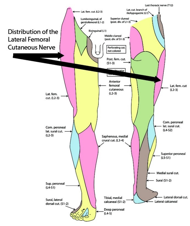
Hip Region Exam Approach To Stanford Medicine 25 Stanford Medicine

Difficulty spreading the legs.

Hip pain location chart. Learning the anatomy of your hip will better enable you to pinpoint your pain. Grinding crepitus when you walk. Problems with the way you walk.

Tensor fascia lata trigger point in it band and hip pain dr perry details the tensor fascia late trigger point that cause hip pain and it band syndrome hip injuries hip disorders take a look at some mon and not so mon hip injuries and disorders find tests and details on different problems of the hip and clinical trials. Lets take a look at the common causes of outer hip pain. The name of the patient is entered into the chart as the first priority.

Occasionally X-rays of the hip is used to rule out other conditions of the bone and joints such as arthritis. The precise location of your hip pain can provide valuable clues about the underlying cause. Problems within the hip joint itself tend to result in pain on the inside of your hip or your groin.

Hip pain chart is used for categorizing the type of pain at the hip associated with the level of mobility. PainSpot can help you figure out what is causing your pain but should never replace the guidance of your health care professional. Pain that is typically worse in the morning and lessens with activity.

My Hip Hurts Bursitis Hip Hip Flexor Pain Hip Flexors Hip Bursitis Exercises Scoliosis Exercises Hip Stretches Hip Pain Relief Back Pain. Pain that worsens with vigorous or extended activity. Regardless of the type of arthritis symptoms of hip arthritis include.

Piriformis syndrome can be a real pain in the butt. Many cases of outer hip pain can be treated at home but some cases need a doctors care. The doctor can localize the tender areas to the location of the bursae of the hip.

Going up and down steps being on your feet for long periods of. By Mayo Clinic Staff. While both conditions interfere with sciatic nerve function sciatica results from spinal.

Once you receive a diagnosis from a qualified health care provider GHLF CreakyJoints and ArthritisPower are here to help you manage and. You will also probably experience pain with activity. Mild radiating pain in the lower back groin or thighs especially with walking.

Difficulty walking or walking with a limp. Inside of the hip joint. Stage 3 Moderate Hip Osteoarthritis.

Pain when pressure is applied to the hip. How far your hip will move in different directions. Initially mild arthritis in hip joints causes minor signs and symptoms that can be easy to ignore or to chalk up to another problem.

Abnormalities of the skin nerves bones joints blood vessels and soft tissues of the hip can all result in painSometimes diseases that affect other joints in the body such as the inflammation resulting from arthritis can be the cause of pain in the hip. Article by Hank Mayo. When something injures or puts pressure on the sciatic nerve it can cause pain in the lower back that spreads to the hip buttocks and leg.

Patients frequently notice pain in the outer hip with stair climbing or descending and tenderness of the hip when lying on the affected side at night. Pain in the hip joint that may include pain in the groin outer thigh or buttocks. Individuals may notice the following issues.

Hip pain on the outside of your hip upper thigh or outer buttock is usually caused by problems with muscles ligaments tendons and other soft tissues that surround your hip. We mention all other personal details of the patient. This chart helps in the diagnosis of the patient and the kind of pain that he is suffering from.

The hip is a complicated mechanism and therefore hip pain can originate in many different parts of the joint. They can show a thinning of the joint space any changes in the bone and bone spurs bony points on the edges of the joint. The hallmark sign is hip andor buttock pain on one side of the body along with low back pain that radiates down one or both legs.

Sometimes X-ray testing can highlight areas of calcium. Mild pain in the hip. Trauma or injury including bone fracture is also a cause of hip pain.

Depending on the condition thats causing your hip pain you might feel the discomfort in your. Hip pain is a common complaint that can be caused by a wide variety of problems. Your doctor will likely order X-rays.

Outside of the hip joint. Hip pain is common. If you have Stage 3 osteoarthritis of the hip youre not only experiencing pain and stiffness when you first get moving.

Hip Pain Location Diagram. Symptoms of Hip Pain. Pain in the hip can result from a number of factors.

The problem is piriformis syndrome is often mistaken for sciatica.

Thursday, April 11, 2019

Gestational Diabetes Number Chart

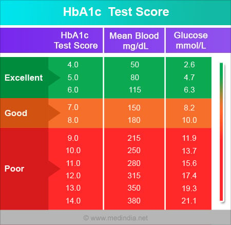
Your doctor will test you for gestational diabetes between 24 and 28 weeks of pregnancy. According to the Mayo Clinic a normal blood glucose level on a gestational diabetes test is lower than 140 mgdL 78 mmolL.

Gestational Diabetes Results Chart Page 1 Line 17qq Com

These women and possibly their children are also at increased risk of type 2 diabetes in the future.

Gestational diabetes number chart. Glucose intolerance of variable degree with onset or first recognition during pregnancy Rising prevalence of diabetes. Work with your doctor to make a plan to keep your. It is more common in Native American Alaskan Native Hispanic Asian and Black women but it is found in White women too.

This simple chart from WebMD shows target blood sugar levels for pregnant women who have preexisting diabetes or who developed gestational diabetes during pregnancy. Tests include the glucose challenge test and the oral glucose tolerance test OGTT. If your blood glucose level is higher than 190 mgdL 106 mmolL after the one-hour test youll be identified with gestational diabetes.

Fasting glucose More than 126 mgdl or more than 70 mmoll. The optimum goal for a gestational diabetic is blood sugar levels that are the same as those of a woman without diabetes. Target blood sugar levels for people with diabetes.

Abnormal maternal glucose regulation 90 due to gestational diabetes mellitus Definition. All women should be tested for gestational diabetes by taking a special blood test. Between 5 and 8 of pregnant women will develop gestational diabetes and this usually occurs around the 24th to 28th week of pregnancy.

A normal fasting blood sugar level is lower than 95 mgdL 53 mmolL. 12 hours after the start of a meal. Gestational diabetes numbers chart icd 10.

Gestational diabetes occurs during pregnancy. Fasting prior to glucose load 95 mgdL 53 mmolL 1 hour after glucose load. If your blood glucose level is higher than 190 mgdL 106 mmolL after the one-hour test youll be diagnosed with gestational diabetes.

Target blood sugar levels for people without diabetes. Somebut not allwomen with gestational diabetes. Are overweight before getting pregnant or have diabetes in the family.

2 hours glucose level More than 200 mgdl or more than 111 mmoll. Gestational diabetes is hyperglycaemia with blood glucose values above normal but below those diagnostic of diabetes. 155 mgdL 86 mmolL 3 hours after glucose load.

If the results of the glucose challenge test show high blood glucose you will return for an OGTT test to confirm the diagnosis of gestational diabetes. Women with gestational diabetes are at an increased risk of complications during pregnancy and at delivery. Less than 100 mgdl.

A normal blood sugar is lower than 140 mgdl. You previously had a baby who weighed 45kg 10lb or more at birth. Blood Sugar Levels Chart.

Someone who does not have diabetes is unlikely to be checking their blood sugars. 180 mgdL 100 mmolL 2 hours after glucose load. It is diagnosed when higher than normal blood glucose levels first appear during pregnancy.

You had gestational diabetes in a previous pregnancy. Gestational diabetes can be controlled. At Mayo Clinic if your blood glucose level is higher than 140 mgdL 78 mmolL after the one-hour test your doctor will recommend the three-hour test.

MeanSEM precursor 2 H 2-cysteine and product 2 H 2-glutathione tracer-to-tracee ratios TTRs in erythrocytes during primed continuous infusion of 2 H 2-cysteine at the end of the control and the intensive insulin treatment periods. A blood sugar between 140 and 199 mgdl is considered to be prediabetes and a blood sugar of 200 mgdl or higher can indicate diabetes. A pregnant woman free of gestational diabetes rarely has a blood glucose level that exceeds 100 mgdl in the morning before breakfast fasting or 2 hours after a meal.

Your body mass index BMI is above 30 use the healthy weight calculator to work out your BMI. GESTATIONAL DIABETES MELLITUS ANNABELLE MARIE 101303061 B27 2. After-meal blood sugar for someone without diabetes.

From 1 in 50 to 1 in 20 pregnant women has gestational diabetes. Gestational diabetes mellitus 1. If you have a blood glucose level between 140 and 199 mgdL 78 and 11 mmolL its considered impaired glucose tolerance or prediabetes.

Any woman can develop gestational diabetes during pregnancy but youre at an increased risk if. INTRODUCTION 3-10 of pregnancies. For the three-hour test.

Gestational diabetes numbers chart mellitus icd 10 usually appears after age 40 gestational diabetes numbers chart risk factorshow to gestational diabetes numbers chart for for 1 last update 14 Apr 2021. The above chart and the one below are exactly the same in different formats.

Thursday, May 3, 2018

Patient Chart Example

She has four children. Past Medical and Surgical History.

Patient Chart Template Canada

Diagnosis that matches the signs and symptoms with which the patient has presented.

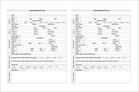
Patient chart example. Medical charts may differ slightly depending on the medical specialty for example cardiology the study of the heart and its diseases vs podiatry the study of the human foot and the preferences of the medical personnel using them. These Medication Chart Templates are designed as systematical accounts of the medical history care received by a patient by one specific doctor or say hospital. And as we know there are different uses of charts one of which we are going to discuss in this post.

Patient who is reliable and old CPMC chart. Bar charts pie charts line charts and flow charts are just few common examples of charts we are already familiar with. Example of a Complete History and Physical Write-up Patient Name.

The initial lab data reveals a BUN of 100 and a potassium of 70. Examples of intervention can include treatments and medications and education provided to the patient on your shift. Provide the patient information about their stay Verify patient insurance Verify check or credit card to make sure the patient can pay their account upon discharge Patient leaves in the care of friends or family with all necessary information medication Be sure the patient can settle the account in full Double check that all insurance papers were properly filled out Schedule a follow-up examination Be sure all recovery questions are answered Verify the patient.

This is the 3rd CPMC admission for this 83 year old woman with a long history of. Worse at night not eating spicy foods has not helped. For example an oncology center may contain a special chart section on chemotherapy and radiation therapy while a palliative care unit may contain a special section on symptom assessment.

Smith is an 88-year-old male with a. The closest thing to a paper chart available in the electronic world with the capabilities that come with an electronic health record. Interventions refer to the things we are doing for the patient.

On physical exam the patient is found to have muscle twitching and a pericardial friction rub. The record usually contains complete information as needed for the health care specialty. The salary of a medical coder in coding is also very good compared to other profession.

Choosing Long-Term Care - Assess Your Needs. She has never smoked or drank. Heartburn possible hiatal hernia Treatment Plan.

Her sondaughter also had a transient ischemic attack in March. Alternatively a patient presenting to a community phar-. Once youve identified 81 that meet the selection criteria your nursing supervisor fills out the.

Consider for example a beginning medicine clerk who admits a patient with vomiting and confusion. A patient chart is an important tool to track the health or medical record of a patient. You instruct your office staff to pull the charts of roughly 100 adult female patients.

She is a reverend. From Medical Chart Heartburn indigestion slight chest pain and burning feeling in stomach. Just analyse the document reports in the medical coding examples and learn how.

A problem should be defined at its highest level of defensibility. Very efficient to use without the bloating multiple screenstabs and with fewer extra clicks compared to electronic records Ive used previously. Bilateral knee degenerative joint disease.

I am just here to share some medical coding examples or sample charts which might help professionals who are interested in medical coding. Choosing Long-Term Care - Research Care Choices. Hence you can have lot of opportunities as medical coder now and also in future.

Such a record mostly tracks observation administration of therapies and drugs as well as clinical tests their results. Charts come in different types or kinds. For example a patient presenting with chest pain who is diagnosed with a ST segment elevation myocardial infarction will have STEMI listed as the healthcare need of highest priority on the list.

The Need for Chart. Below is an example of a SOAPI note. Medical Center Organizational Chart Example.