The most common mechanisms of injury are either a fall that causes a direct blow to the greater trochanter or forced lateral rotation of the lower extremity Bucholz et al 2009. A 77-year-old woman height 150 cm body weight 43 kg with osteoporosis associated with high bone turnover was treated with oral cyclical etidronate 400 mgday for 2 weeks every 3 months.
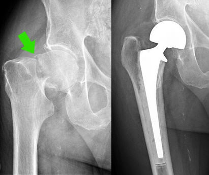 Osteoporosis Case Study 1 Hip Fracture Of The Femoral Neck
In a nutshell osteoporosis neck pain does not exist.

Femoral neck osteoporosis. Acase of insufficiency fracture of the femoral neck that occurred during treatment for osteoporosis is reported. Does Osteoporosis Neck Pain Exist. The femoral neck which is located near the top of the femur bone is especially susceptible to fractures due to osteoporosis because it is the weakest part of the femur.
The type of surgery however depends on the severity and location of your particular hip fracture. The T-score reference value being derived from bone density measurements in a population of young healthy females see table and figure below diagnosis of osteoporosis relies on its operational definition which is when an individuals T-score for BMD at the femoral neck is equal to or more than 25 SDs below the reference value Kanis JA Diagnosis of osteoporosis and assessment of fracture risk. The WHO Osteoporosis Working Group and other international osteoporosis organizations have stated that the femoral neck is the only site that should be used in the estimation of osteoporosis prevalence at a population level.
The femoral neck is a frequent site of osteoporosis in elderly women after their menopause. I often recommend the squat as a femoral neck osteoporosis exercise. Osteoporosis is a pervasive disease among the growing elderly population.
The ROC curve showed the poor diagnostic value of the Singh index since the area under the curve was approximately 40 the Singh index is therefore a poor screening tool for femoral neck osteoporosis. The femoral neck which is located near the top of the femur bone is especially susceptible to fractures due to osteoporosis because it is the weakest part of the femur. Lets review proper squat form.
Trouble twisting or bending your body and pain when you do. Can cause bone pain but the neck would be an unusual presentation. However the high impact activities are not suitable for people with severe osteoporosis.
Though this is because of the chemical imbalance cause by cessation of certain hormones it is propagated further by a sedentary lifestyle. The neck that they see in their DEXA BMD results is actually the neck of the femur or femoral neck not the neck on their shoulders that supports their head. Sudden severe back pain that gets worse when you are standing or walking with some relief when you lie down.
To identify a pathology is difficult because it is not diagnosed by a bone density test a diagnostic procedure designed to determine the degree of osteoporosis. Osteoporosis refers to a decrease in the calcium content of bones which makes them brittle and weak. The squat assuming your knees are able to handle it is probably the best femoral neck osteoporosis exercise you can do.
Pathology associated with impaired bone mineralization. Osteopenia at the femoral neck observed in old age. 3 4 However an objective of the present study was to estimate the number of people with osteoporosis and low bone mass as defined by the clinical guidelines from the NOF which are based on bone status at either the femoral neck.
They do have neck pain but it is not a result of their osteoporosis. Femoral neck fractures occur most commonly in the eighth decade of life as a result of bone that is weakened by either osteoporosis or osteomalacia. If you fracture your hip youll probably need surgery.
Femoral neck fractures are often a direct result of osteoporosis and are challenging to treat. My view is that exercises done in a vertical position ie. She is a precipitating factor of fractures of the femur.
A clinical diagnosis of osteoporosis may also be established without bone mineral density measurement by the presence of a fragility fracture particularly at typical sites spine hip pelvis wrist humerus or rib. Surgical interventions seek to return the patient to preinjury function as quickly as possible but many obstacles exist. The condition can lead to weakening of the hip bone.
There are two main types of hip fracturesfemoral neck fractures and intertrochanteric region fractures. Resistance exercises targeting the femoral neck and the surrounding muscles and connective tissues can help strengthen the bone and thereby decrease your risk for fractures. Jumping running and to a lesser extent jogging and walking are the best exercises for improving femoral neck BMD.
Femoral neck osteopenia refers to a decrease in bone density in the top part of the femur where it connects to the hip explains the University of Maryland. Resistance exercises targeting the femoral neck and the surrounding muscles and connective tissues can help strengthen the bone and thereby decrease your risk for fractures. Ideally with time and as you feel strong and as your form is solidified you want to eventually move to doing weighted squats.
The reference standard site of bone mineral density analysis is the femoral neck but other sites such as lumbar spine can be used. Femoral Neck Fractures These fractures occur where your femur fits into your hip joint.