Showing posts with label photos. Show all posts

Saturday, September 4, 2021

Inflammatory Breast Cancer Photos

These photos are frank they were taken before diagnosis during chemotherapy just before mastectomy surgery radiation markings immediately post radiation 16 days post radiation and final healing of chest wall. Although it is often a type of invasive ductal carcinoma it differs from other types of breast cancer in its symptoms outlook and treatment.

Breast Cancer Gynecology And Obstetrics Merck Manuals Professional Edition

Cancer that starts in the breast.

Inflammatory breast cancer photos. Inflammatory breast cancer is a rare and very aggressive type of breast cancer that tends to spread quickly even in the relatively early-stagesInterestingly the signs and symptoms of IBC are quite common. Most cases are invasive ductal carcinomas which develop in the cells lining the milk ducts and spread. Inflammatory breast cancer IBC is a rare and aggressive malignancy that is often initially misdiagnosed due to its similar presentation to more benign breast pathology such as mastitis resulting in treatment delays.

One of the most common malignancies in women in the US Breast cancer malignant breast neoplasm is cancer originating from breast tissue most commonly from the inner lining of milk ducts or the lobules that supply the ducts with milk. Remember there are often non-visual symptoms that include itching pain and skin thickening. Studies have found that women with inflammatory breast cancer who are treated with a multimodal approach have better responses to therapy and longer survival.

Inflammatory breast cancer is an infrequent aggressive type of breast cancer that spreads rapidly. This video aired in 2007 and yet when I shared it with my close friends none of them had ever heard of it. 13 May 2020 1045 in response to graymackay Hi all I hope you are all ok Im the same I had breast cancer in 2013 a year after I lost my husband to cancer so was quite a shock when I was diagnosed it was luckily caught early so had lumpectomy radiotherapy and tamoxifen which I stopped.

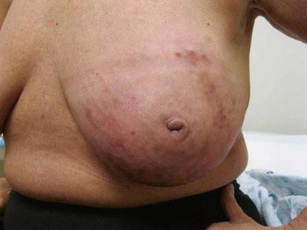
Its not pretty but its critical information. Accounting for one to five percent of all breast cancer cases in the United States inflammatory breast cancer or IBC is an aggressive rare form of this disease. You can see how red the breast is and the square-ish mass of thickened skin as well as the peau dorange skin.

Inflammatory breast cancer IBC is a rare and aggressive form of breast cancer. The lymph vessels found in skin of the breast. 4 rows Inflammatory breast cancer includes many stages which can be tested.

So few people have heard of this type of cancer. The bottom pic was taken 14 days after first chemo. Inflammatory breast cancer IBC is rare and accounts for only 1-5 of all breast cancers.

Thank you to the IBC patients who have provided these images for use on this web site. After your staging tests are. However these signs and symptoms are most often associated with benign breast conditions.

Check Inflammatory Breast Cancer Pictures images to examine itchy rash bruises red spots discoloration or pain in breasts with early signs symptoms. In 2020 an estimated 276480 new cases of invasive breast cancer are expected to be diagnosed in women in the US. Extreme swelling that can pop up almost overnight with inflammatory breast cancer.

Inflammatory breast cancer is generally treated first with systemic chemotherapy to help shrink the tumor then with surgery to remove the tumor followed by radiation therapyThis approach to treatment is called a multimodal approach. Cancer initiates when normal cells in the breast alter and grow uncontrollably forming a sheet of cells called a tumor. Skin dimpling can be a sign of inflammatory breast cancer because cancer cells.

About 1 in 8 US women about 12 will develop invasive breast cancer over the course of her lifetime according to the American Cancer Society. The Inflammatory Breast Cancer Research Foundation wishes to thank her for sharing her story and photos with the IBC community. Inflammatory breast cancer.

Breast cancer cancer of the breast. Inflammatory breast cancer is a rare and aggressive form of breast cancer that occurs when malignant cells block the skin and lymph vessels of the breast. You may not associate breast cancer with redness or a skin rash but in the case of inflammatory breast cancer IBC a rash is an early symptomThis is an aggressive form of breast cancer.

This video shows photos of inflammatory breast cancer. Inflammatory breast cancer pictures show a red andor swollen breast that appears inflamed. Inflammatory breast cancer usually starts with the reddening and swelling of the breast instead of a distinct lump.

Jo urn al Pr e-p roo f Figure 1 Caption Pictures of two patients with inflammatory breast cancer are shown. Most instances are in the cells lining the milk ducts and spread through the entire breast invasive ductal carcinomas which grow. Top 2 pictures are prior the first chemo.

Inflammatory Breast Cancer Pictures. Inflammatory Breast Cancer Research Foundation These photographs show typical visual clinical symptoms that appear at time of diagnosis before treatment. Inflammatory breast cancer images reveal andor a reddish bloated breast that appears inflamed.

According to the American Cancer Society about 1 of all breast cancer cases in the United States are inflammatory breast cancers. The usual cause of inflammatory breast symptoms is breast mastitis or duct ectasia.

Wednesday, July 7, 2021

Face Cancer Photos

This picture gallery contains some examples of the more common. It is commonly found on sun-exposed skin on the face forearms andor hands.

The Spot Under This Woman S Eye Looked Like A Pimple Mdash But It Was Skin Cancer Health Com

663 for regional.

Face cancer photos. This warning sign may indicate an invasive BCC. Skin cancers can look very different. Skin Cancer Pictures by Type.

Melanoma a serious form of skin cancer is often curable if you find it early. Often the first sign of melanoma is a change in the shape color size or feel of an existing mole. April 18 2011 507 PM CBS News.

Previous Next 1 of 6 Melanoma pictures for self-examination. Squamous Cell Carcinoma SCC Up next on this roundup of skin cancer pictures. But this common form of cancer can also occur on areas of your skin not ordinarily exposed to sunlight.

Skin cancer is the most common form of cancer. Squamous cell carcinomas often look like scaly red or skin-colored patches open sores warts or elevated growths with a central depression and may crust or bleed. Is it skin cancer.

A spot or sore. Skin Cancer on Scalp Pictures - 20 Photos Images Skin cancer the abnormal growth of skin cells most often develops on skin exposed to the sun. These skin cancer pictures will help you differentiate the different types including basal cell and squamous cell carcinomas melanoma actinic keratosis and merkel cell carcinoma.

Other signs such as diameter and evolution are not shown as they are difficult to assess through static images. The beginning signs of skin cancer involve a change in the skin. Based on data from 2010 to 2016 the 5-year relative survival rates for melanoma of the skin in the United States are 927 percent overall and.

The American Academy of Dermatology advises that you watch skin spots for these features. Science Photo Library - DR. There are several different types of skin cancer with Basal Cell Carcinoma Squamous Cell Carcinoma Bowens Disease Keratoacanthoma Actinic Keratosis and Melanoma most commonly occurring.

There are many types of skin cancer each of which can look different on the skin. Squamous cell carcinoma the second most common form of skin cancer. Melanoma is a type of cancer that begins in melanocytes cells that make the pigment melaninBelow are photos of melanoma that formed on the skin.

Nodular basal cell carcinoma is responsible for 60-80 of. The skin appears shiny and taut often with poorly defined borders. More than 60000 people are diagnosed with melanoma every year with nearly 9000 deaths according to Skin Cancer Foundation data.

Melanoma can also start in the eye the intestines or other areas of the body with pigmented tissues. Tawny Willoughby a 27-year-old nurse who grew up in a small Kentucky town shared a photo to Facebook in April ahead of Mays Skin Cancer Awareness Month. Nodular basal cell carcinoma is a type of skin cancer that is most often found on the head.

Since not all BCCs have the same appearance these images serve as a general reference to. Skin Cancer Image Gallery. Photos of skin cancer.

A scar-like area that is flat white yellow or waxy in color. However when you do a self-examination of your skin make. Early Signs of Skin Cancer Pictures - 15 Photos Images.

Skin cancer is by far the most common type of cancer. The skin cancer pictures give you an idea of what skin cancer can look like. Many pathologists consider it to be a form of squamous cell carcinoma the second most common type of skin cancer.

99 for localized melanoma. I recommend that you store these images and this answer to bring them with you to the dermatologist to confirm the diagnosis and discuss treatment options. So its important you visit your GP as soon as possible if you notice a change in your skin.

This type of cancer starts in basal cells which are tasked with making new skin cells to push the old ones toward the surface of the skin. MARAZZI Getty Images. Basal cell carcinoma is the most common form of skin cancer and least dangerous whereas melanoma often referred to as.

These signs are shown below each picture. Nearly all skin cancers can be treated effectively if they are found early so knowing what to look for is important. 38 photos that could save your life PICTURES By David W Freeman.

Signs of skin cancer differ in form color and borders. This may mean that a new lump or sore has formed on the skin that a new mole has popped up or that an existing mole has begun to grow or change in shape. Itchy crusty or bleeding.

Actini Keratosis AK is a pre-cancerous growth. The earlier a skin cancer is diagnosed the easier it is to treat. These melanoma pictures can help you determine what to look for.

Melanoma is the deadliest type of skin cancer. A red or dark patch. The image -- which is below -- shows her face after using a cream called Aldara or imiquimod to treat skin cancerThe cream is used in lieu of surgery to treat superficial basal cell carcinoma.

Saturday, April 7, 2018

C Diff Stool Photos

See clostridium difficile stock video clips. C diff Stool Appearance What does C diff look like.

Clostridium Difficile C Diff Symptoms Causes Treatment

Bowel movements that contain blood and mucus are concerning and may be indicative of more severe C diff disease.

C diff stool photos. The c diff stool appearance when you have c diff diarrhea is usually watery has a mucus like consistency like nasal discharge when you have a bad cold with shreds of solid matter in the flow. What does c diff diarrhea look like. C diff stool photos The c diff stool appearance when you have c diff diarrhea is usually watery has a mucus like consistency like nasal discharge when you have a bad cold with shreds of solid matter in the flow.

The c diff stool appearance when you have c diff diarrhea is usually watery has a mucus like consistency like nasal discharge when you have a bad cold with shreds of solid matter in the flow. Huge collection amazing choice 100 million high quality affordable RF and RM images. Clostridium difficile bacterium Clostridium difficile bacterium isolated on black background 3D illustration.

Read Janes Story To Learn A Solution To See Better Nails Within Two Weeks. C diff diarrhea will have a very strong odor that differs from typical diarrhea. Read Janes Story To Learn A Solution To See Better Nails Within Two Weeks.

A significant prolonged increase in the amount of mucus in the stool is a potential sign of potential disorders such as Crohns disease cystic fibrosis irritable bowel syndrome or a malabsorption issue such as celiac disease or lactose intolerance. Emuaid Defeats Severe Nail Fungus. In mild cases you may have to go to the toilet for more than 3 times a day however in severe cases you may need to go for 10-15 times a day.

Ad I Found One Fast Simple Trick. Bacteria which cause pseudomembraneous colitis and are associated with nosocomial antibiotic resistance clostridium difficile stock pictures royalty-free photos images. Ad I Found One Fast Simple Trick.

In controlled laboratory settings however where the nurses were blind to patient characteristics presence of diarrhea antibiotic use. No need to register buy now. Your stool will return to its normal color once you are eating regularly and stop tajing antibiotics.

Here are some pictures of C. Click here for Clostridium difficile pictures. Emuaid Defeats Severe Nail Fungus.

Bismuth subsalicylate the active ingredient in many upset stomach medications can change the color of your stools to. The burgundy color may indicate blood in the stool. Base on the bristol stool chart below C diff looks like number 6.

Other studies have shown that nurses can accurately identify C Diff stools by odor in up to 84 sensitivity and 77 specificity. You can also find pictures of Mycoplasma pneumonia Enterovirus Lunginflammation. Find the perfect c diff stock photo.

Find the perfect clostridium difficile stock photo. 6 wks post cdiff solid stool tested for toxin a b eia test came back negative. 307 clostridium difficile stock photos vectors and illustrations are available royalty-free.

No watery d for 7 weeks. C diff diarrhea is usually watery and rarely bloody. Glad to see you are on the way to recovery.

Gram-positive Clostridium difficile bacteria revealed in the stool sample micrograph film 2004. Photos of c diff poop. Other conditions can cause this so be aware that only a c diff test can confirm that you are excreting c diff.

No need to register buy now. Sem of clostridium difficile bacteria - clostridium difficile stock pictures royalty-free photos images Clostridium difficile seen under optical microscopy. I am finishing flagly for c-diff.

28 de fevereiro de 2021 Sem categoria. Huge collection amazing choice 100 million high quality affordable RF and RM images. 2 You should let your doctor know about the presence of increased mucus pus or blood in your stool.

Pictures c difficile stools. Diff infections can cause stomach pain diarrhea fever and loss of appetite. Other conditions can cause this so be aware that only a c diff test can confirm that you are excreting c diff toxin.

Normal value ranges might differ somewhat among various labs. Clostridium clostridium difficile infection bacteria resistant to antibiotics bacteria spore food bacteria nosocomial anaerobic infections. Difficile infection can cause watery stools both in mild cases and severe cases.

What does C diff Smell Like.